Theroy Preparation-0 (Part 2)

1️⃣ *PSEUDOLYMPHOMA*

2️⃣ PITYRIASIS LICHENOIDES

Febrile ulceronecrotic Mucha Habermann disease👇🏻

3️⃣ PARAPSORIASIS

4️⃣ LYMPHOCYTOMA CUTIS

5️⃣ JESSNER’S LYMPHOCYTIC INFILTRATE

Epidermal melanin unit

Distribution of melanocytes

Melanoblast migration and  differentiation

Melanosome transport

Melanosome transfer to keratinocytes

Melanocyte culture

Biochemistry of melanogenesis

Regulation of human pigmentation

Melanocyte regulation by endocrine factors

Melanocyte regulation by paracrine and autocrine

Biological significance of melanin

Constitutive pigmentation

Classification of disorders of melanin pigmentation

Physiological Hypermelanosis

Facial melanosis

Melasma

Photocontact melanosis

Poikiloderma of Civatte

Erythromelanosis follicularis of the face and neck

Peribuccal pigmentation of Brocq

Ephelides

Lentiginosis

🔰 Hypermelanosis due to endocrine disorders

1️⃣ Addison disease

2️⃣ Acromegaly

3️⃣ Cushing syndrome

4️⃣ ACTH administration

5️⃣ Hyperthyroidism

🔰 *Hyperpigmentation in other systemic disorders*

🔰 Neoplastic disease

Hyperpigmentation in rheumatoid disease

Systemic sclerosis and morphea

SLE and dermatomyositis

Neurological disorders

Multiple organ failure

Renal failure

Primary biliary cirrhosis

Hemochromatosis

Cutaneous amyloidosis

Nutritional deficiency

POEMS syndrome

🔰 Hypermelanosis of drug origin

✅ Very important👆🏻

Fixed drug eruption

Pigmentation resulting from acute photodynamic and phototoxic reactions

Post inflammatory hypermelanosis

Ashy dermatosis and erythema dyschromicum perstans

Treatment of hypermelanosis

1️⃣ Vitiligo

2️⃣ Halo naevus

🔰 Acquired syndromic hypomelanosis

1️⃣  Vogt- koyanagi- Harada syndrome

2️⃣ Alezzandrini syndrome

Post inflammatory hypomelanosis

Chemical depigmentation

Idiopathic guttate hypomelanosis

Punctate leukoderma

Endogenous non melanin pigmentation

Cutaneous hemosiderosis

Jaundice and bronze baby syndrome

Carotenoderma

Ochronosis

🔰 Important tip for this chap.👇🏻

Dun waste too much time on reading this chap from rooks.

Just listen to above voice notes and solve mcqs

Happy learning😊

🔰 Table 91.1 in rooks 9th edition is a summary of the whole topic Rosacea.

🔰 Summary of rosacea👆🏻

ETTR

Pathophysiology👇🏻

Pathology👇🏻

Causative organisms 👇🏻

Demodex mite

Clinical features

Clinical variants 👇🏻

Granulomatous rosacea

Differential diagnosis👇🏻

Only read this table for DDs

Complications👇🏻

Disease course and prognosis👇🏻

Investigations

Management👇🏻

Only revise tables for management👆🏻

🔰 *Important note*👇🏻


For Rosacea, only listen to above voice notes, revise tables from rooks and practice mcqs.

Its more than enough for this chap

Associations 👇🏻

Pathophysiology👇🏻

Pathology👇🏻

Diagnostic criteria👇🏻

All 3 must be fulfilled

Clinical features👇🏻

Classification of severity👇🏻

 *Hurley staging system*

Hurley stage 1

Hurley stage 2

Hurley stage 3

Differential diagnosis👇🏻

Complications 👇🏻

Disease course and prognosis👇🏻

Investigations 👇🏻

Management👇🏻

🔰 *Important note*👇🏻


For HS, above voice notes are enough. Reading from rooks can be done for self satisfaction.

Practice from mcqs later on.

🔰 Disorders of pilosebaceous unit and disorders of sweat glands can be covered from mcqs.

Dun waste ua time reading it from rooks.

Clubbing👇🏻👇🏻

Koilonychia

Pincer nail

Nail shedding

Onycholysis

Pterygium

Longitudinal grooves👇🏻

Transverse grooves and beau’s lines

Trachyonychia

Onychoschizia/ lamellar dystrophy

Nail plate pigmentation

Subungual disturbances

Nail bed changes

Leuconychia

Colour changes due to drugs and chemicals

YELLOW NAIL SYNDROME

Red lanulae

Longitudinal erythronychia

Splinter hemorrhage

🔰 *Causes of Splinter hemorrhage*


Liver disease

Endocarditis

Connective tissue disease

Meningococcal disease

Psittacosis

Disseminated histoplasmosis.


 *Skin disease*


 psoriatic nail disease

lichen planus 


 *Systemic diseases*


 microemboli or injury to vessel walls associated with vasculitis

Primary antiphospholipid syndrome


Systemic lupus erythematosus


Raynaud disease


Behcet disease


Cutaneous vasculitis


Scurvy.


chronic kidney disease on haemodialysis or post-renal transplant


 *Drugs*


Tyrosine kinase inhibitors (seen in 60–70% of patients taking sunitinib and sorafenib)


Nitrofurantoin


Ganciclovir


Terbinafine


Tetracyclines.

Mneumonic of painful tumours

7 types

Superficial

Dystrophic

Proximal

Endonyx

Paronychia

Mixed

Dlso

Nail examination by worthy Dr Saadat sb👆🏻

📝 Hutchinson eye sign …in herpes zoster ..

Hutchinson nail sign.. melanoma.. ( others are pseudo Hutchinson and micro Hutchinson)

Hutchinson triad in congenital syphilis ..

Hutchinson summer prurigo which is actinic prurigo.

Hutchinson malignant freckle which is lentigo maligna

What is the significance of this… Black streak…

 *Melanonychia*


Here there can be the following *possibilities*


 *1.functionsl melanonychia* ( melanocyte activation,common in dark individuals, can be due to trauma ,)


 *2.melanocytic nevi*


 *3.inflammatory skin conditions* like lp and other pigmentary disorders which can be correlated clinically)

 The pigmentation varies from brown dark brown to black

Pseudo Hutchinson can be seen too.


 Most commonly involves thumb but other nails can be involved as well


 *_Mostly a naevi  in matrix !*_

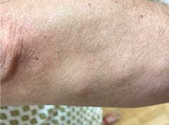
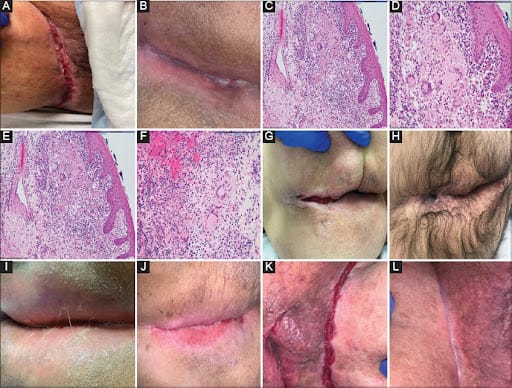
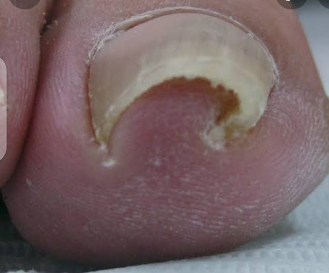
mostly these are *matrix naevi*  leading to band, so focal matrix ablation might lead a nail plate dystophy lateron which might look uglier than this


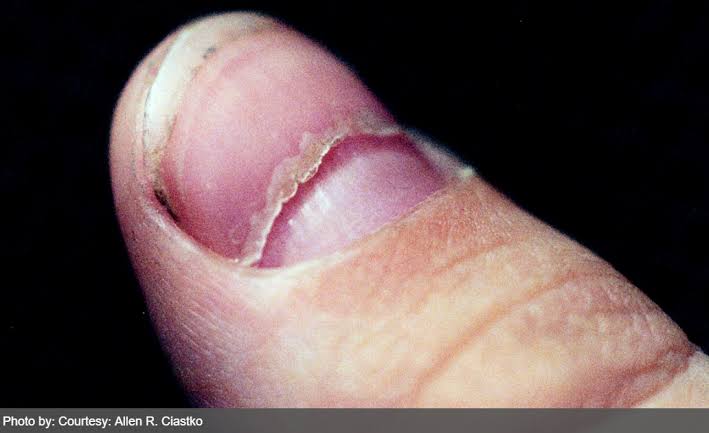
Would the nail appearance be normal after this procedure and complete healing?


 *Yes only the transverse lenght* *will be shortened* , rest it will be perfact


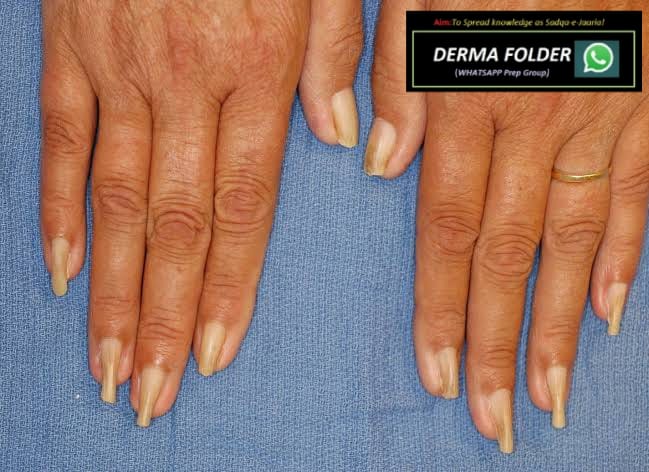
Grabbing this opportunity to revise *ABCDEFGHI*  , to R/O *subungual melanoma* in such cases


*A: Age (* 50–70 years old); African, Japanese, Chinese, and Native American heritage


 *B: Brown-black* pigmented band ≥3mm with blurred borders


 *C: Change or lack of change* despite treatment in the nail band or nail morphology


 *D: Digit most* commonly involved (thumb, big toe, or index finger)


 *E: Extension* of pigment into the skin surrounding the nail (Hutchinson sign)


 *F: Family or personal history* of melanoma or dysplastic nevus (atypical mole)


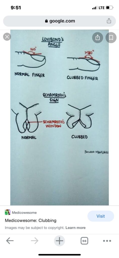
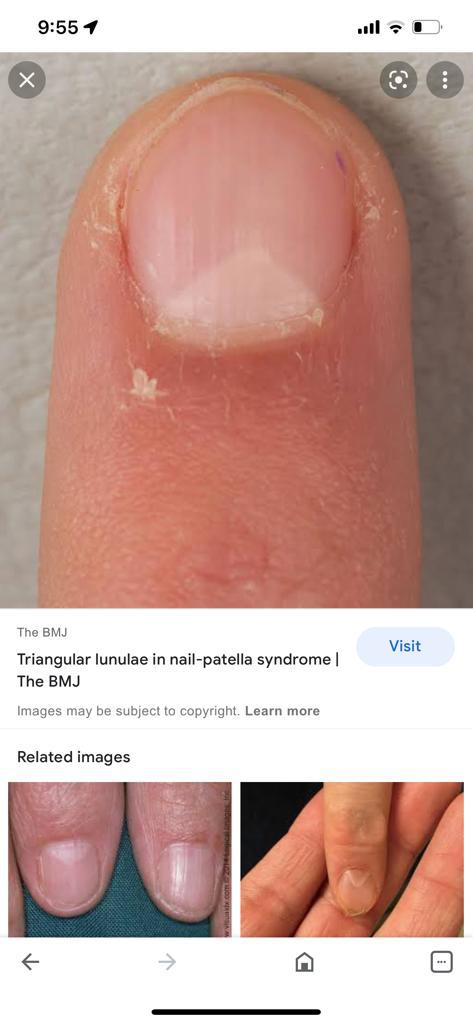
 *G – Geometry* of lesion like triangular band


 *H – Hyponychial involvement*


 *I – irregular pigment pattern* (like different color bands) , irregular spaces , irregular thickness
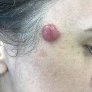
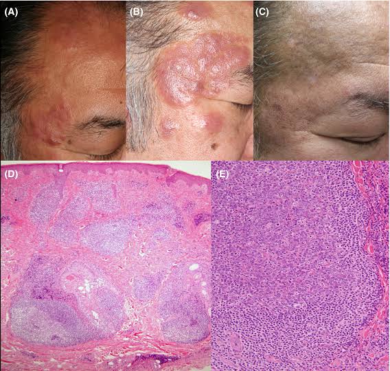
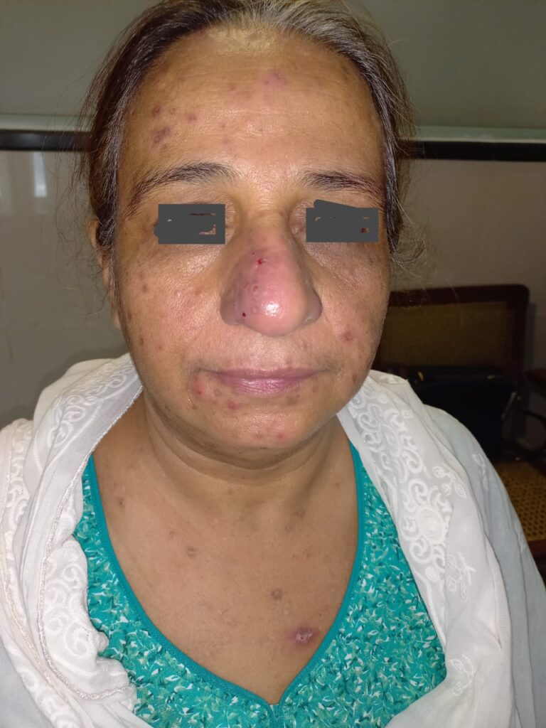
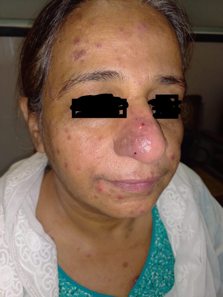
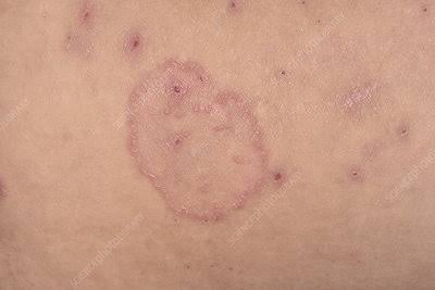
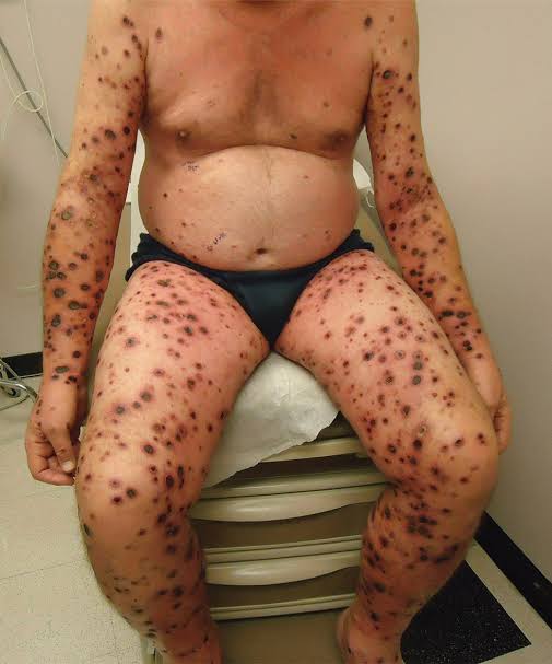
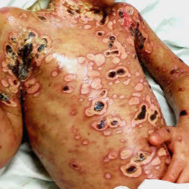
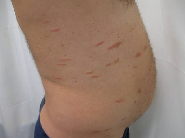
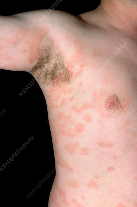
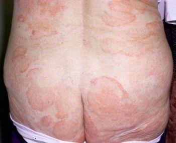
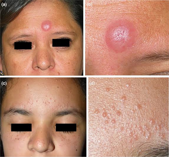
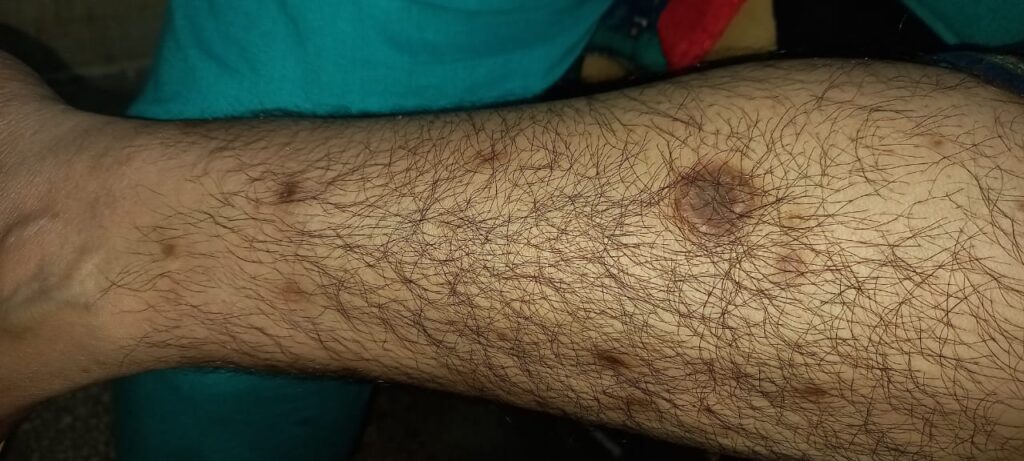
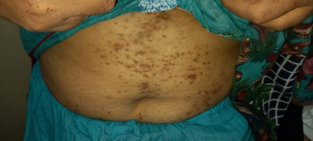
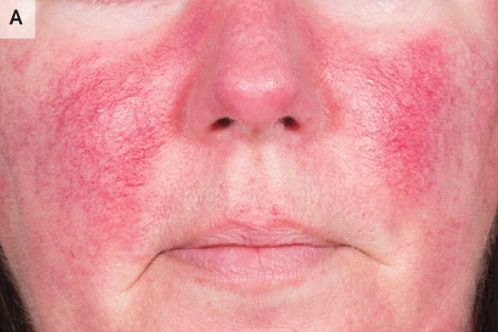
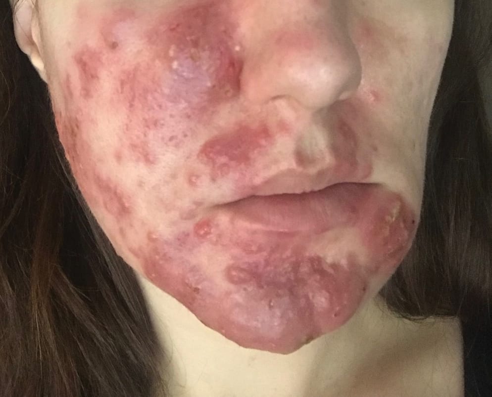
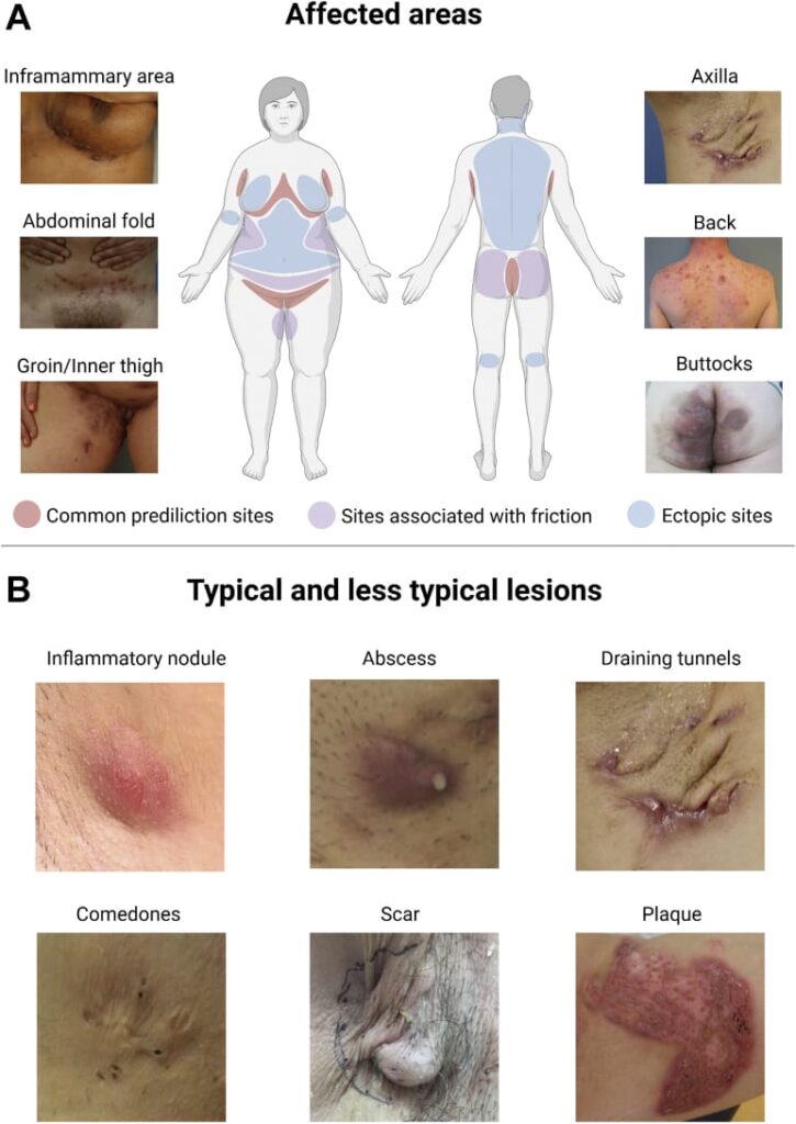
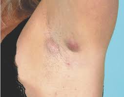
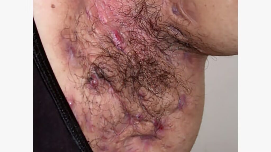
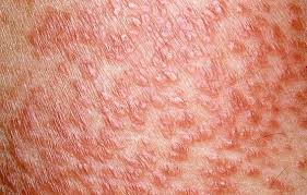
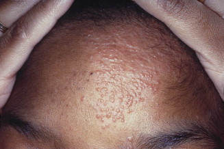
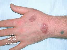
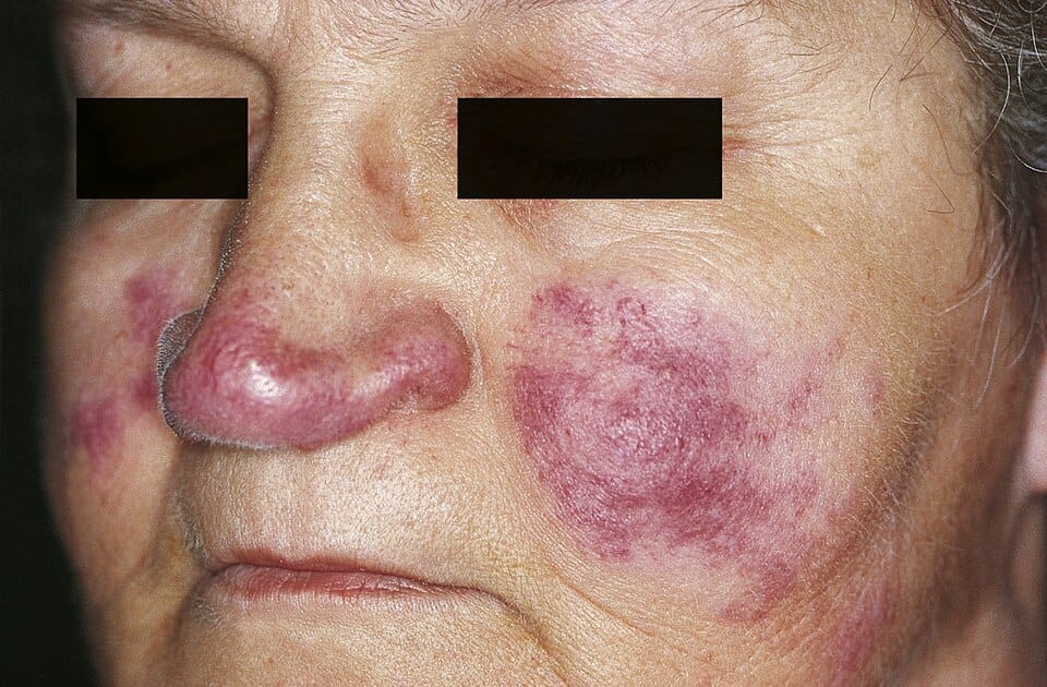
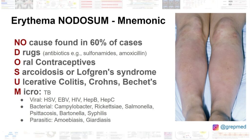
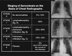
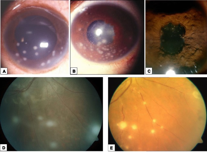
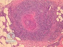
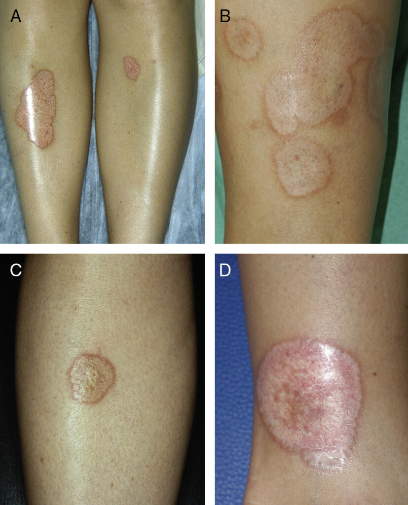
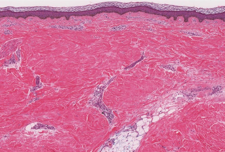
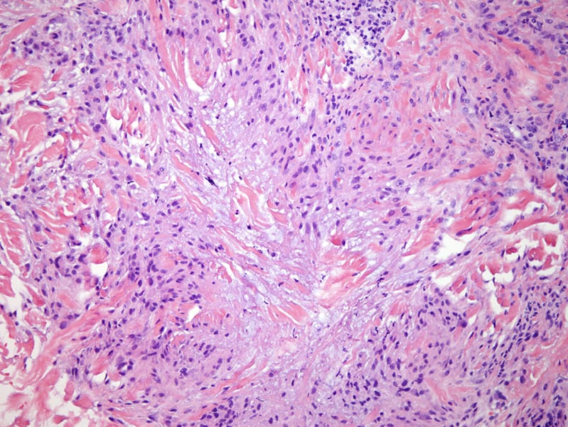
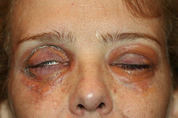
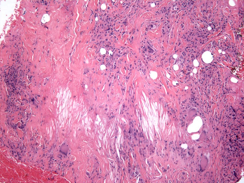
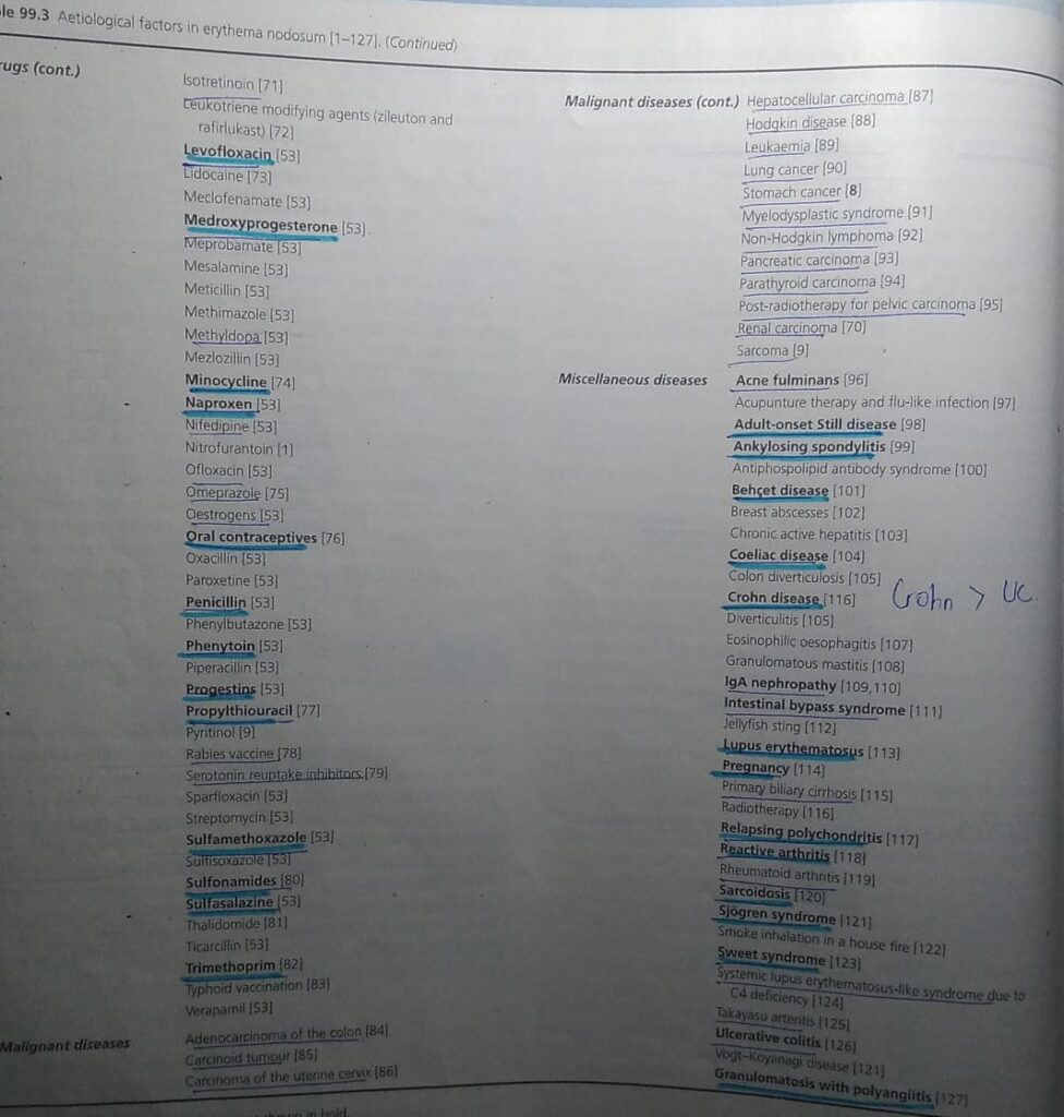
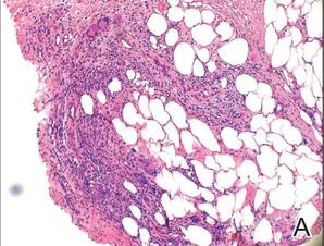
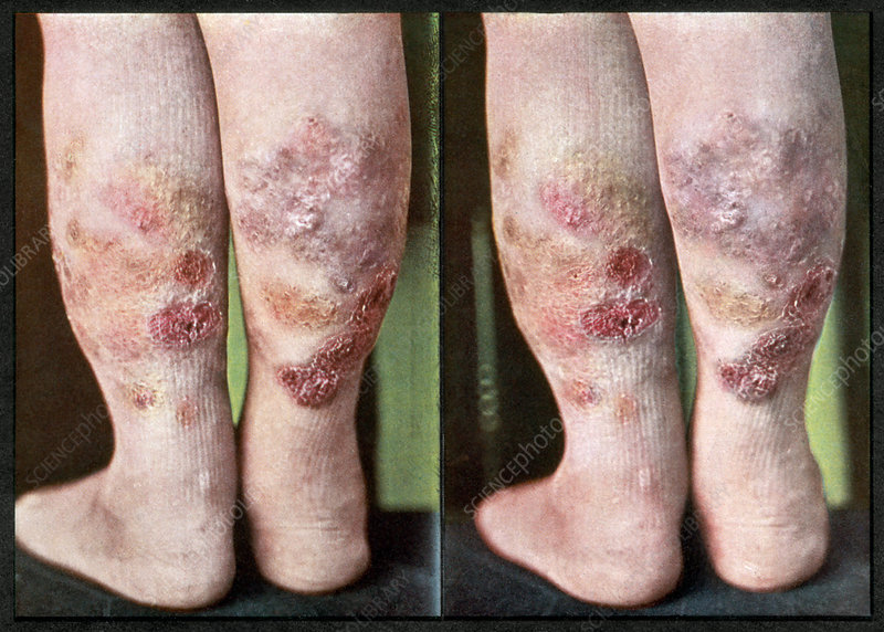
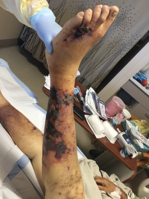
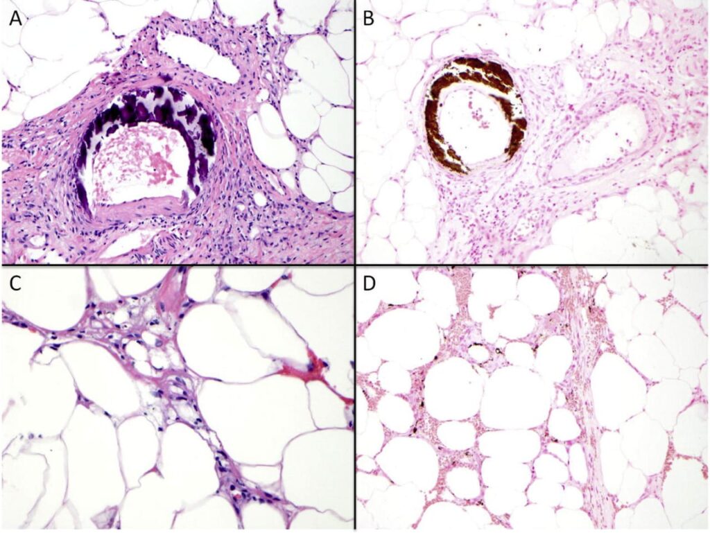
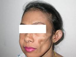
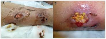
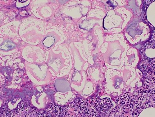
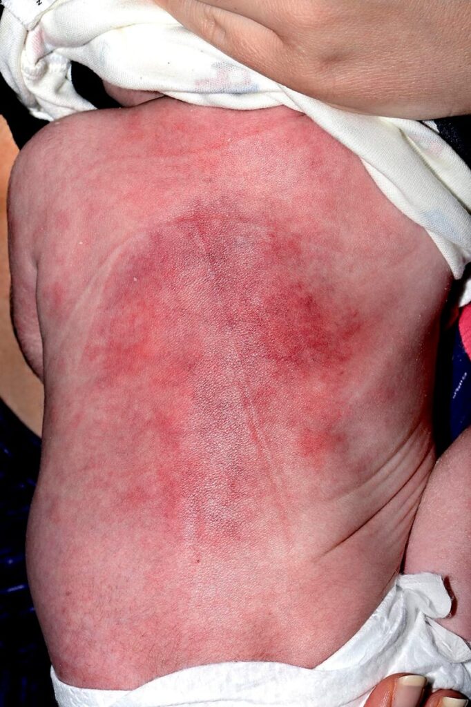
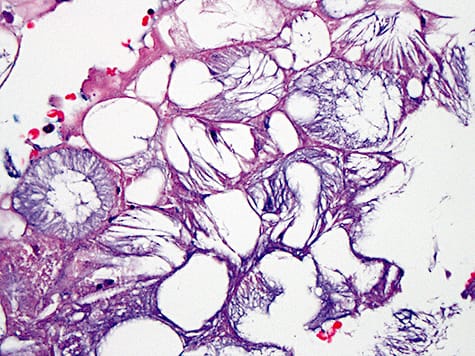
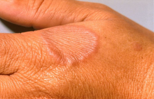
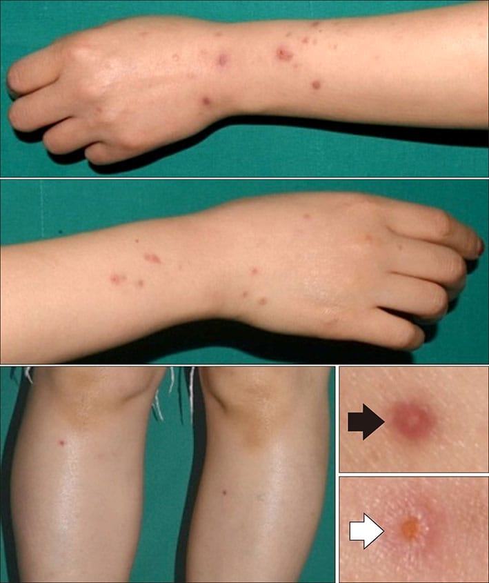
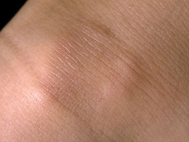
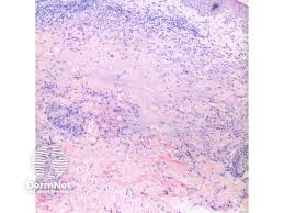
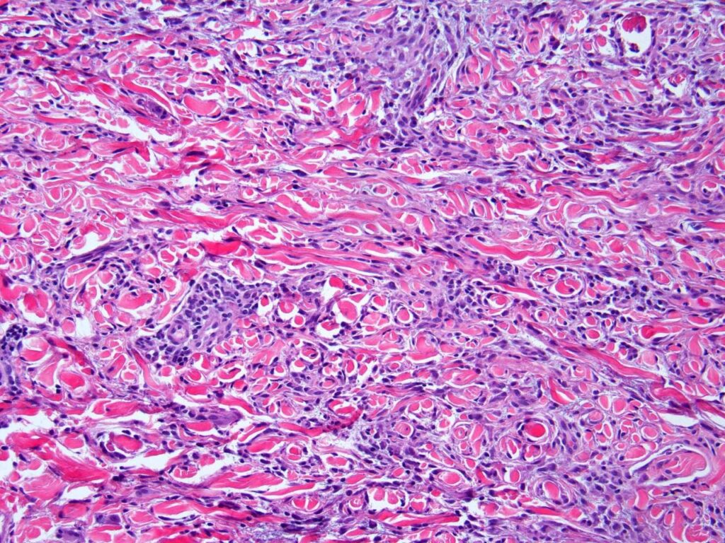
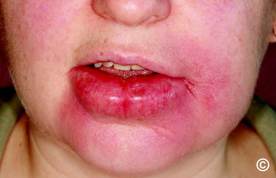
🔑 🗝 🔐 Pincer nails deformity


*hereditary disease*


▪️autosomal dominant Mendelian

▪️Clouston


 *acquired pincer nails*


▪️gastrointestinal malignancies,

▪️renal failure,

▪️Kawasaki disease,

▪️amyotrophic lateral sclerosis, and

▪️systemic lupus erythematosus

🔰 Racquet nails

🟥 *causes?* 🔑


*Genetic disorders*


▪️Larsen syndrome, 

▪️Brooke–Spiegler syndrome, 

▪️Rubinstein–Taybi syndrome, 

▪️Hajdu–Cheney syndrome, 

▪️cartilage–hair hypoplasia, 

▪️pycnodysostosis, 

▪️acrodysostosis,

▪️brachydactyly type D.


 *Acquired racquet nail*

associated with 

▪️acroosteolysis and 

▪️psoriatic arthropathy.

▪️diagnostic of bone resorption in hyperparathyroidism.

Anonychia

🔰 Important tip for this chap👇🏻

Pathogenesis👇🏻

Pathology👇🏻

Basophilic fibrinous sheath

🔰 Imp tip for histopathology👆🏻

Differential diagnosis 👇🏻

Triggers👇🏻

Clinical features👇🏻

1️⃣ Cutaneous sarcoidosis

🔯 Specific forms of Cutaneous sarcoidosis

✅Maculopapular sarcoidosis

Typical appearance of lesions of sarcoidosis

✅ Nodular and Plaque sarcoidosis

✅ Lupus pernio

✅ Scar sarcoidosis

Scar sarcoidosis

✅ Subcutaneous sarcoidosis

Sarcoid dactylitis

🔯 Less common forms of cutaneous sarcoidosis

Heerfordt syndrome 👇🏻

Heerfordt syndrome

Mneumonic

Mikulicz syndrome 👇🏻

2️⃣ Pulmonary sarcoidosis

3️⃣ Ocular sarcoidosis

4️⃣ Reticuloendothelial system

5️ Liver

6️⃣ Neurosarcoidosis

7️⃣ Others

🔰 Course and prognosis 👇🏻

🔰 Investigations 👇🏻

🔰 Management

🔰 *IMPORTANT TIP*


Above voice notes covers all imp aspects of Sarcoidosis.

So if u skip reading it from the book and only listen to these voice notes + practice with mcqs will be sufficient.

🔰 IMPORTANT TIP FOR THIS CHAP👇🏻

Septal Panniculitis

Basophilic fibrinous sheath

Imp points to remember👆🏻

Stains used in biopsy specimens of Panniculitis

🔰 SEPTAL PANNICULITIS

1️⃣ Cutaneous PAN👇🏻

2️⃣ Necrobiosis lipoidica 👇🏻

3️⃣ Deep morphea👇🏻

4️⃣ Subcutaneous granuloma annulare👇🏻

5️⃣ Rheumatoid nodule👇🏻

6️⃣ Necrobiotic xanthogranuloma👇🏻

7️⃣ *Erythema nodosum*👇🏻

Zoom in

1️⃣ Erythema nodosum leprosum 👇🏻

2️⃣ Erythema induratum of Bazin

2️⃣ Erythema induratum of Bazin

3️⃣ Calciphylaxis👇🏻

4️⃣ Cold panniculitis

Horse rider’s pernio

5️⃣ *Lupus panniculitis/ lupus erythematosus profundus*

6️⃣ Pancreatic panniculitis👇🏻

7️⃣ Alpha-1 antitrypsin deficiency panniculitis

8️⃣ Infective panniculitis

🔰 *Important mneumonic* 👇🏻


Ghost adipocytes can be seen in pancreatic panniculitis, mucormycosis and aspergillosis


MAP – mneumonic

Follow the map to reach the ghost

9️⃣ *Subcutaneous fat necrosis of newborn*

🔟 Sclerema neonatorum👇🏻

🔰 *IMPORTANT TIP*


Panniculitis is an imp chap for theory and viva exam.

They imp topics and tips mentioned here are the most useful and now onwards u l never find this chap difficult in sha Allah.

Simply remember the table in the beginning of the chapter and imp histopath findings

Topics which arent covered are less likely asked but must practice them from mcqs

Happy learning😊

1️⃣ Granuloma annulare

*Clinical features*👇🏻

Localized granuloma annulare

Generalized GA

Perforating GA

Subcutaneous GA

Histopathology👇🏻

  1. Necrobiotic palisading granulomas
  2. Interstitial form
  3. Granulomas of tuberculoid or sarcoidal type

*Differential diagnosis*👇🏻

Complications

*Disease course and prognosis*

Benign self resolving lesions

40% patients may show recurrence

*Investigations*

*Management*

*Drugs causing abnormal platelet function*👇🏻


  1. NSAIDs
  2. Penicillin and beta lactam antibiotics
  3. Prostacyclin and iloprost
  4. Alteplase and other fibrinolytic drugs
  5. Ticlopidine and clopidogrel
  6. Glycoprotein IIb/IIIa antagonist
  7. CCBs, nitrates, quinidine
  8. Streptokinase

🔰 *NON THROMBOCYTOPENIC VASCULAR CAUSES OF PURPURA* 👇🏻

🔯 *RAISED INTRAVASCULAR PRESSURE*

1️⃣ *Gravitational purpura*

2️⃣ *Acroangiodermatitis*

3️⃣ *Exercise induced purpura*

🔰 *IMPORTANT NOTE*

For this chap, listen to above notes and solve mcqs for practice.