Theroy Preparation-0 (Part 1)

8️⃣ Calcifying aponeurotic fibroma

9️⃣ Dermatomyofibroma 👇🏻✅✅

Plaque like CD-34 positive dermal fibroma 👇🏻

Angiomyofibroblastoma

Cellular angiofibroma 👇🏻

Elastofibroma

Inclusion body (digital) fibromatosis 👇🏻✅

Fibroma of tendon sheath 👇🏻

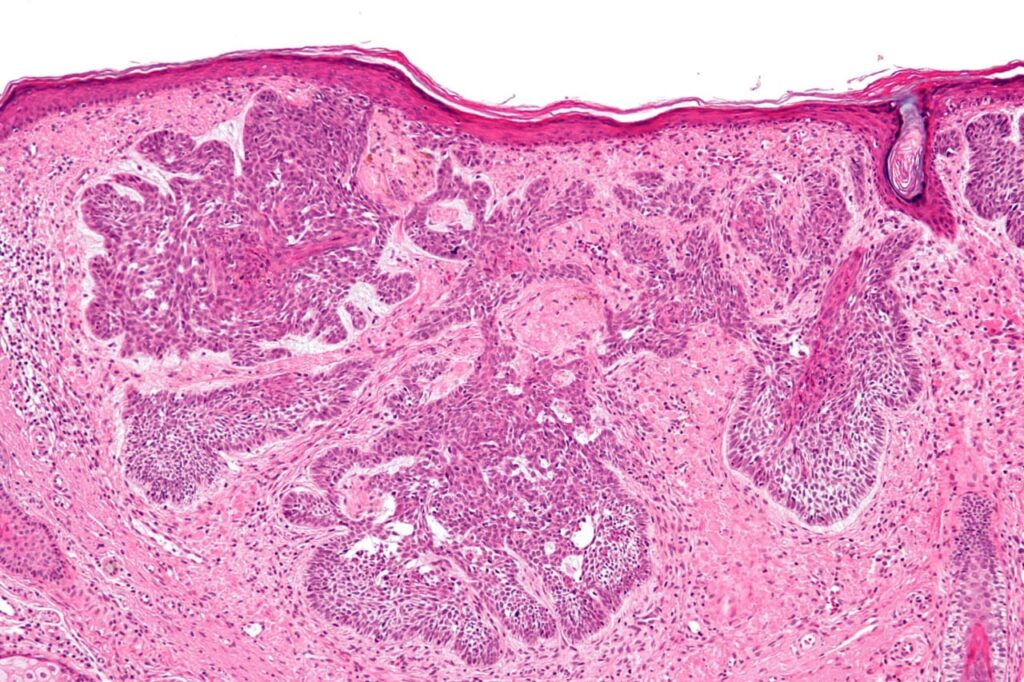
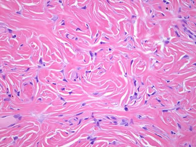
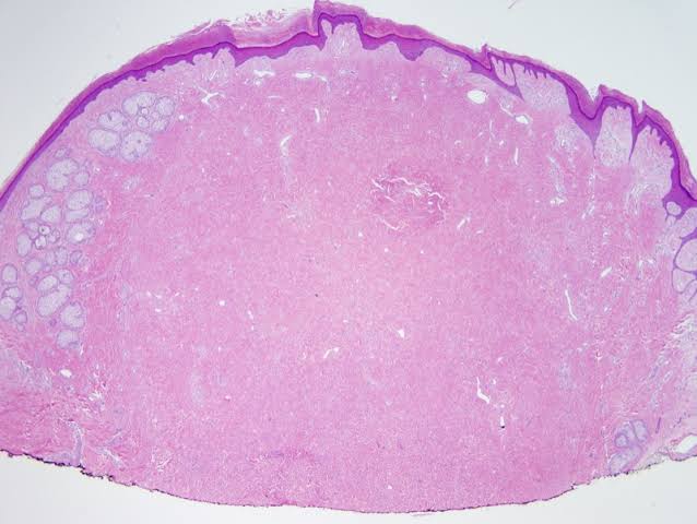
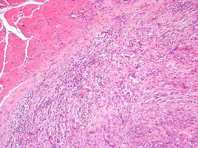
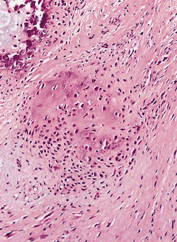
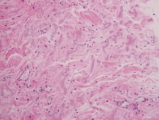
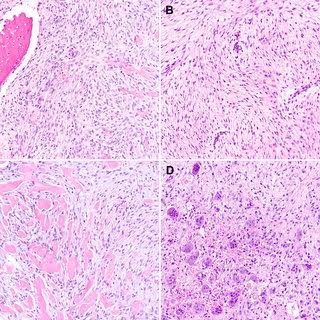
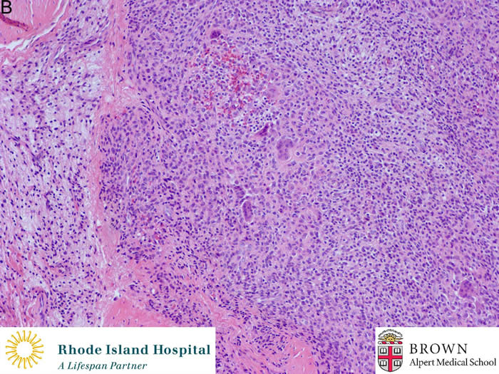
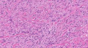
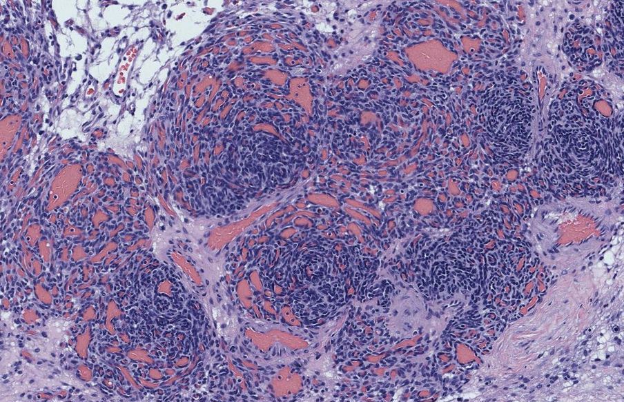
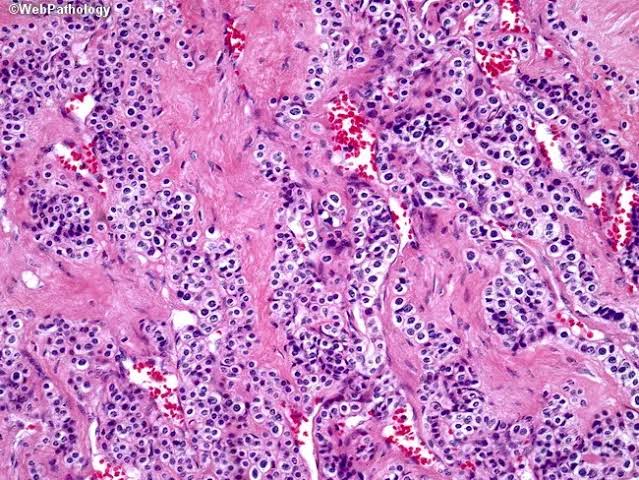
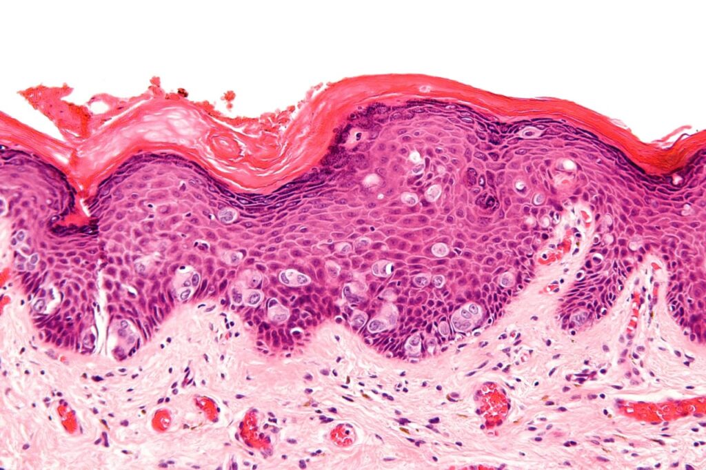
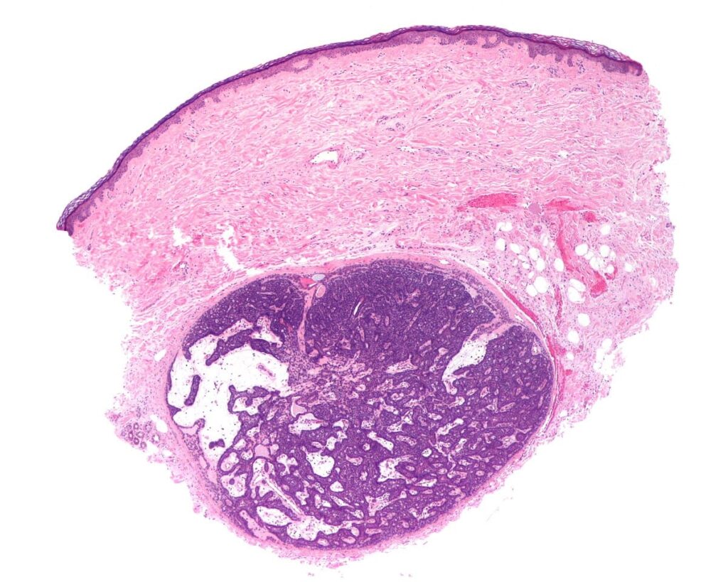
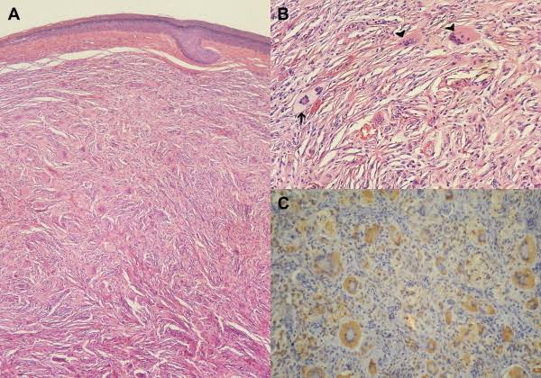
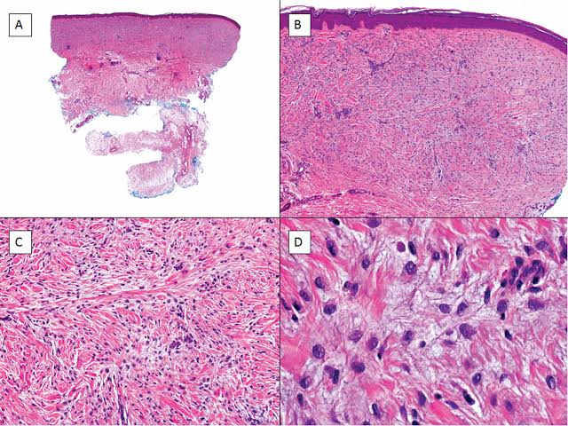
Fibroma of tendon sheath is well-circumscribed and has a lobular growth pattern like giant cell tumor of tendon sheath. The degree of cellularity is variable. There are hypocellular areas with sparse numbers of bland spindle cells in a dense collagenous stroma adjacent to zones of myxoid change containing stellate cells.

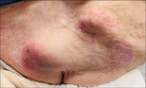
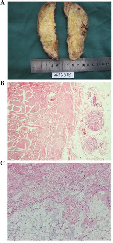
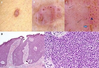
Desmoplastic fibroblastoma 👇🏻

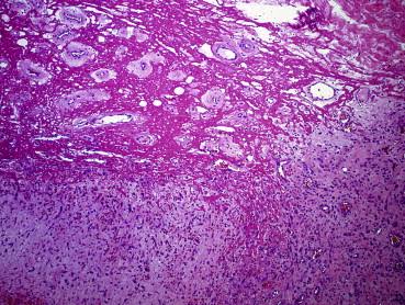
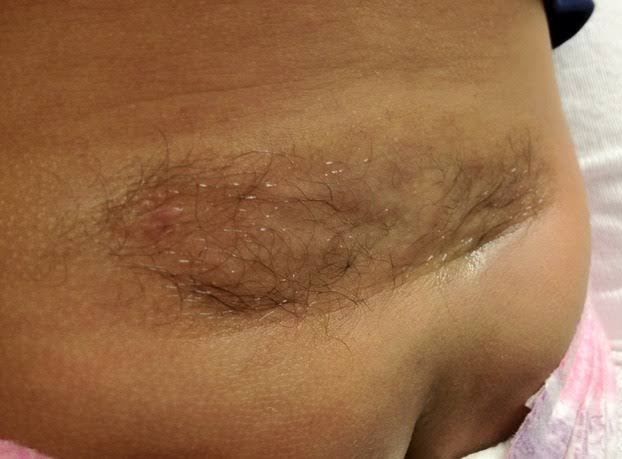
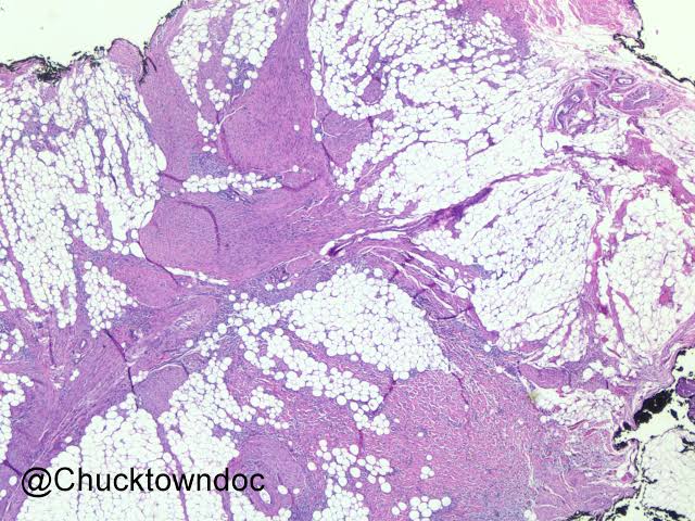
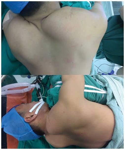
Nuchal type fibroma 👇🏻

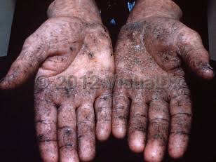
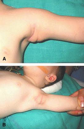
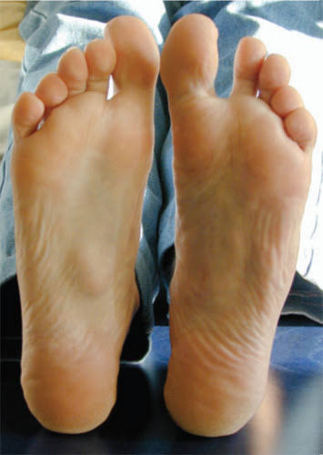
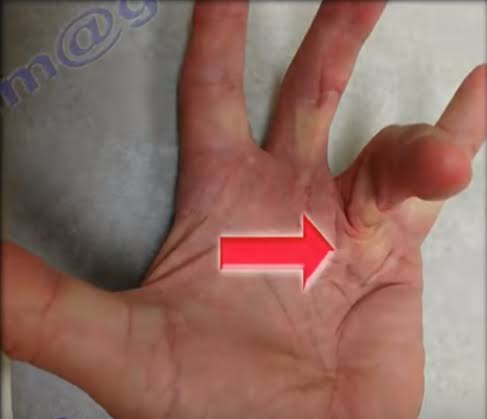
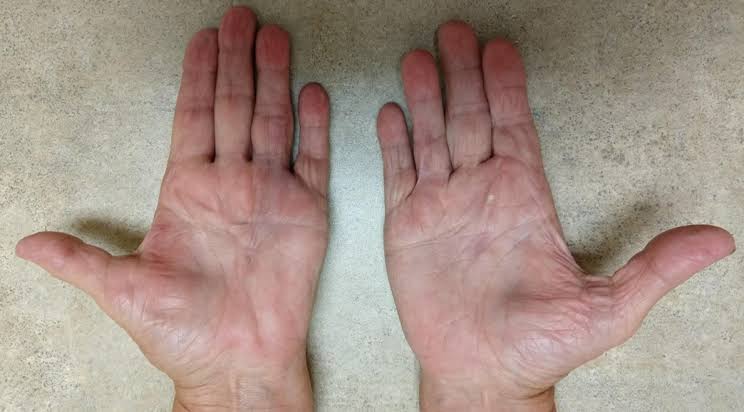
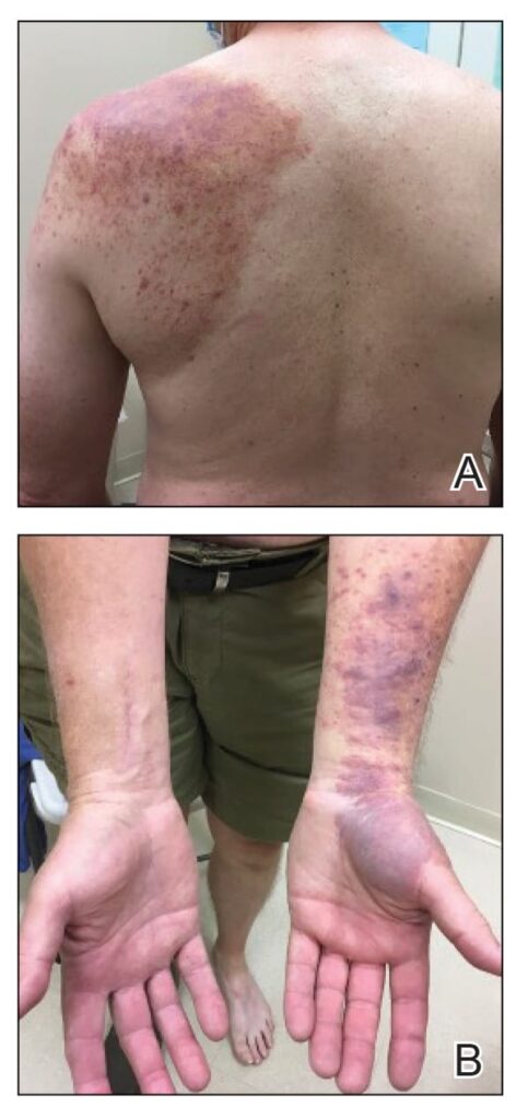
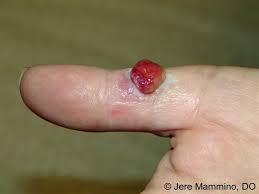
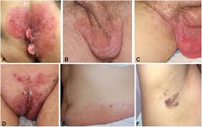
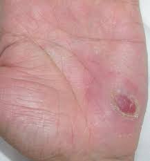
Palmar and plantar fibromatosis (superficial fibromatosis) 👇🏻✅

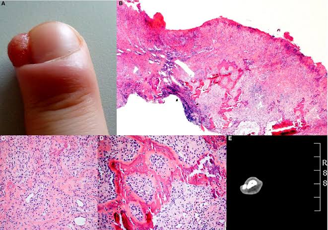
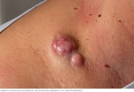
Penile fibromatosis 👇🏻✅

🔰 *Important note*

Soft tissue tumours is one of the imp chaps of rooks which usually comes in mcq exam and in opds as well.

Its difficult to memorize bcoz for diagnosis histopath is must to remember.

Thts y if any such questions comes in exam u can only diagnose it if u know the histopath of tht particular tumour or its IHC markers.

Dermatofibrosarcoma protuberans✅✅

🔰 This image came in our toacs exam

3️⃣ Pyogenic granuloma / Lobular capillary hemangioma✅

4️⃣ Kaposiform haemangioendothelioma✅

5️⃣ Angiosarcoma

Spiradenoma✅

A very imp exam slide

Cylindroma✅

Mixed tumour of skin

Hidradenoma

Eccrine hydrocystoma

Syringoma✅

Apocrine carcinoma

🔰 *IMPORTANT NOTE*


For this chap, u must memorize the histopathology of imp topics which frequently comes in exam(which i have marked imp).

ETAS will help u to make the concept clear.

In the end, u ll only remember the points mentioned or explained in mcqs so spend more time in mcqs

Clinical features

ABCDE

Diagnostic tools

SUPERFICIAL SPREADING MELANONA

NODULAR MELANOMA

LENTIGO MALIGNA MELANOMA

ACRAL LENTIGINOUS MELANOMA

SUBUNGUAL MELANOMA

MUCOSAL MELANOMA

OCULAR MELANOMA

AMELANOTIC MELANOMA

REGRESSIVE MELANOMA

MALIGNANT BLUE NAEVUS

SINONASAL MELANOMA

Strategies for early detection

Investigations

HISTOPATHOLOGICAL DIAGNOSIS

Immunohistochemistry

Prognostic markers

Biopsy

Staging of Primary melanoma

Follow up

Wide local excision of melanoma

Sentinel lymph node biopsy

Systemic treatment of melanoma

Systemic therapy for metastatic disease

Advanced stage IIIB/IIIC and stage IV disease

The mitogen-activated protein kinase (MAPK) pathway includes cascades of protein kinases which are activated by genotoxic stress and growth factors, including chemotherapeutic compounds. MAPK pathway activation stimulates the RAS oncogene, which stimulates RAF, consequently leading to ERK1/ERK2 cascade.

Chemotherapy

Summary of systemic rx

🔰 IMPORTANT TIP 👇🏻

🔰 DISORDERS OF DENTRITIC CELLS

1️⃣ Langerhans cell histiocytosis 👇🏻

(Most imp topic of this chap)

For revision go through tables

🔰 Revise Management from the  Treatment ladder

🔰 Dendritic cell origin

👇🏻Disorders with mainly skin involvement

1️⃣ Juvenile xanthogranuloma

Touton giant cell is imp to remember along with markers

2️⃣ Benign cephalic histiocytosis

3️⃣ Generalized eruptive histiocytosis

4️⃣ Papular xanthoma

5️⃣ Progressive nodular histiocytosis

6️⃣ Xanthoma disseminatum

7️⃣ Diffuse plane xanthomatosis

👇🏻Disorders of skin with predominant skin involvement

1️⃣ Erdheim-Chester disease

Predominant skin involvement with/without systemic component👇🏻

1️⃣ Reticulohistiocytoma

2️⃣ Familial sea blue histiocytosis

3️⃣ Hereditary progressive mucinous histiocytosis

4️⃣ Malakoplakia

5️⃣ Necrobiosis xanthogranuloma

👇🏻Disorders with predominant systemic involvement and some skin involvement

1️⃣ Multicentric reticulohistiocytosis

🔰1 of the most imp topics of this chap. Very imp for theory and clinical slides. Imp differential too👆🏻

2️⃣ *Sinus histiocytosis with massive lymphadenopathy/ Rosai Dorfman disease*

1. Benign keratinocytic acanthomas and proliferation

2. Cutaneous cysts

Solve mcqs only

🔰 Mycosis fungoides

🔰 *Important tip*


MF is the most imp topic of this chap, before exam this topic is a must to revise.

Make ua concept from above voice notes and then revise MF from long case group which is  more refined and summarized.

Solve mcqs from various sources to make the concept even more clear.

Imp parts are clinical variants, markers, clinical presentation, staging, histopath and management.


Happy learning😊

Histopath of MF👆🏻

Sezary syndrome

🔰 All tables given in rooks in the section of MF are v imp.

They ll help u with quick revision

1️⃣ *Intravascular large B cell lymphoma*

2️⃣ *Lymphomatoid granulomatosis*

Cutaneous manifestations of hodgkin’s disease

3️⃣ *Primary cutaneous B cell lymphomas*

4️⃣ *Marginal zone lymphoma*

5️⃣ *Follicle centre cell lymphoma*

Diffuse large cell lymphoma

*Recent advances in MF* treatment include:

    – Histone deacetylase inhibitors (HDACi) like romidepsin and belinostat

    – Immune checkpoint inhibitors like pembrolizumab and nivolumab

    – Brentuximab vedotin (an antibody-drug conjugate)

    – Mogamulizumab (a monoclonal antibody)

    – Lenalidomide (an immunomodulatory agent

– zonalimunab (anti CD 4 antibody)

– alemtuzumab (anti CD 52)

💥MUCOSAL MELANOTIC LESIONS

👇🏻Pigmented melanotic macules

💥DERMAL MELANOCYTIC LESIONS

👇🏻Naevus of Ota

👇🏻Naevus of Ito

💥CONGENITAL MELANOCYTIC NAEVUS

👇🏻Speckled lentiginous naevus

💥 ACQUIRED MELANOCYTIC NAEVI

Naevus spilus👆🏻

Compound naevus👆🏻

Intradermal naevus👆🏻

💥 NAEVI IN UNUSUAL SITES

Melanocytic naevi of genital area

Acral naevi

Conjunctival naevi

Naevi of nail matrix or nail bed

💥 NAEVI WITH UNUSUAL MORPHOLOGY

Meyerson naevus

Cockade naevus

Targetoid haemosiderotic naevus

💥SPITZ NAEVUS

🔰 Histopath of spitz naevus is an important exam slide.

Atypical spitz naevus

Blue naevus and variants

🔰 Important tip for this chap👇🏻


For the identification of the type of a naevus, it is v imp tht u understand and remember its histopathology, so u must have a gud grip over it.

Its an imp exam slide too, both for slides and toacs station so prep it very well.

Happy learning😊